Bone and Soft Tissue Neoplasms

Share this post
Case 12: Typical Distal Femur Lesion
www.bstneoplasms.com
Appendicular Bone Lesions

Case 12: Typical Distal Femur Lesion

31-years old with knee pain

Bhavin Jankharia
Mar 15
Share this post
Case 12: Typical Distal Femur Lesion
www.bstneoplasms.com

If you would like to know immediately, each time a new case is up via email, please subscribe.

A 31-years old presented with knee pain.

The X-rays (simulated from the CT scan) showed a distal femur lesion. This was followed by MRI and then a biopsy.

X-rays simulated from CT scan
MRI

Histopath

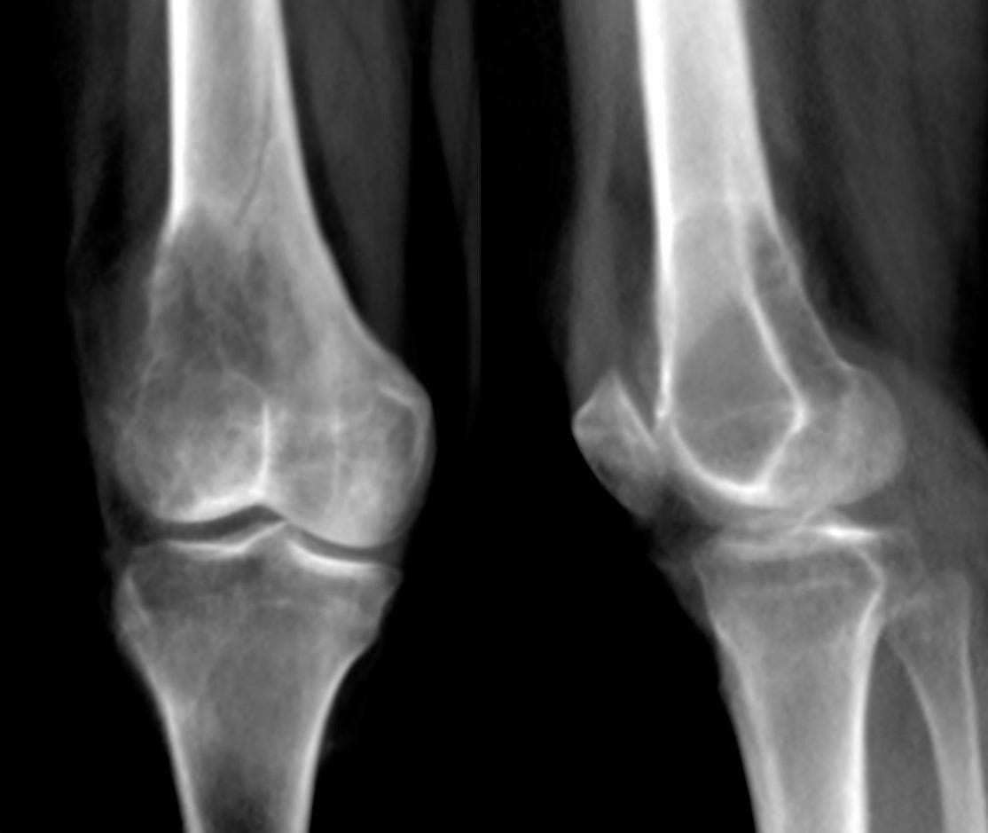
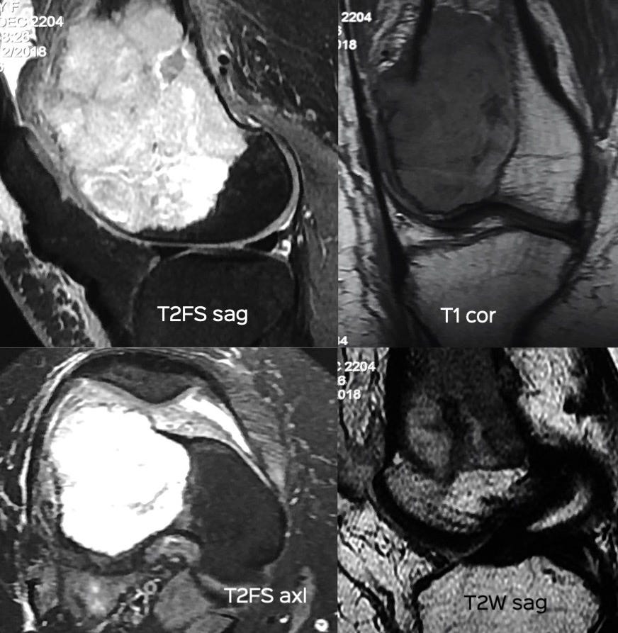
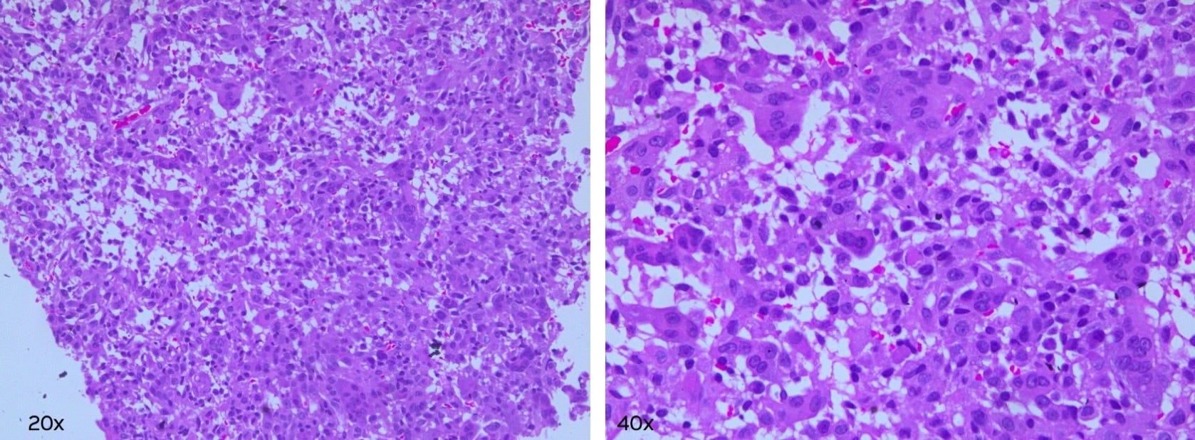
What is the most likely diagnosis?

  1. Giant cell tumor

  2. Chondroblastoma

  3. Osteosarcoma

  4. Aneurysmal bone cyst

The answer is up here.

Please share with those who you think might benefit from this post / case.

Share


Other Cases You May Like

https://www.ctbiopsy.com/angulation03/

https://www.atmasvasth.com/happiness/

https://www.ctchestreview.com/ctr/

Share this post
Case 12: Typical Distal Femur Lesion
www.bstneoplasms.com
Comments

Create your profile

0 subscriptions will be displayed on your profile (edit)

Skip for now

Only paid subscribers can comment on this post

Already a paid subscriber? Sign in

Check your email

For your security, we need to re-authenticate you.

Click the link we sent to , or click here to sign in.

TopNewCommunity

No posts

Ready for more?

© 2022 Bhavin Jankharia
Privacy ∙ Terms ∙ Collection notice
Publish on Substack Get the app
Substack is the home for great writing