
Case 65: Mono-articular Knee Arthropathy
48-years old with progressive knee pain

Case:
48 years old had progressive knee pain.
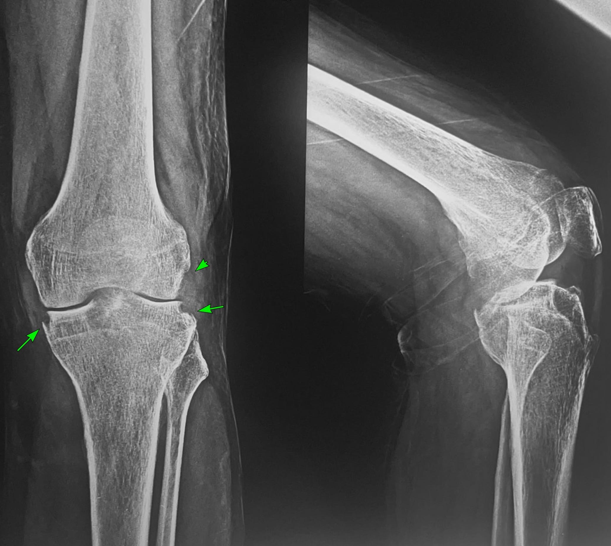
Radiograph (Fig. 1) shows large articular margin erosions (arrows) along the peripheral margins with mild periarticular osteopenia.
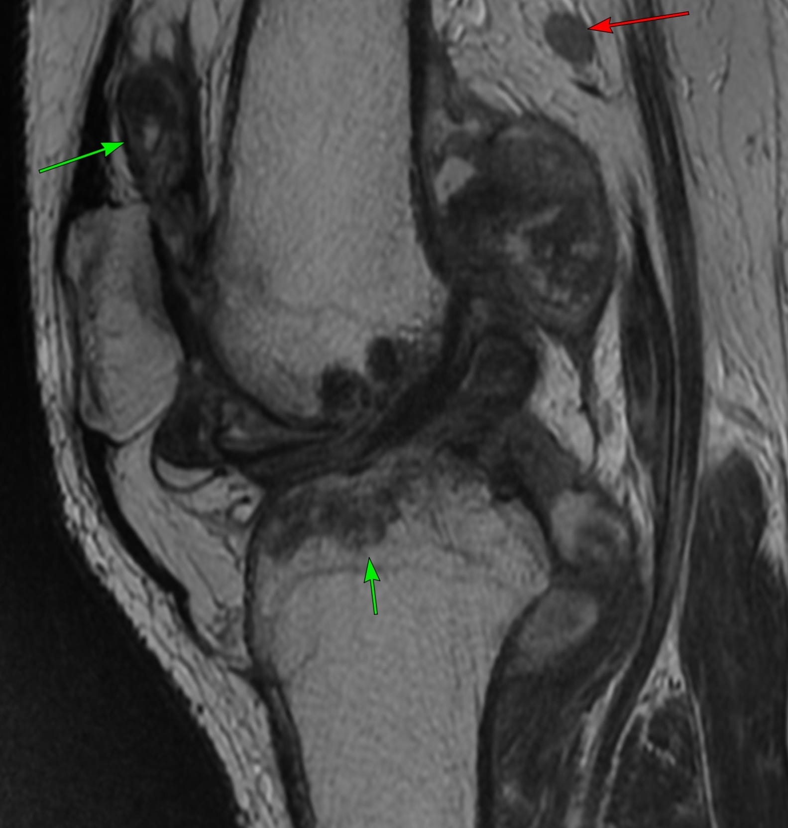
The T2 sagittal image (Fig. 2) shows large synovial nodules (arrows), T2 dark with an enlarged popliteal node (red arrow).
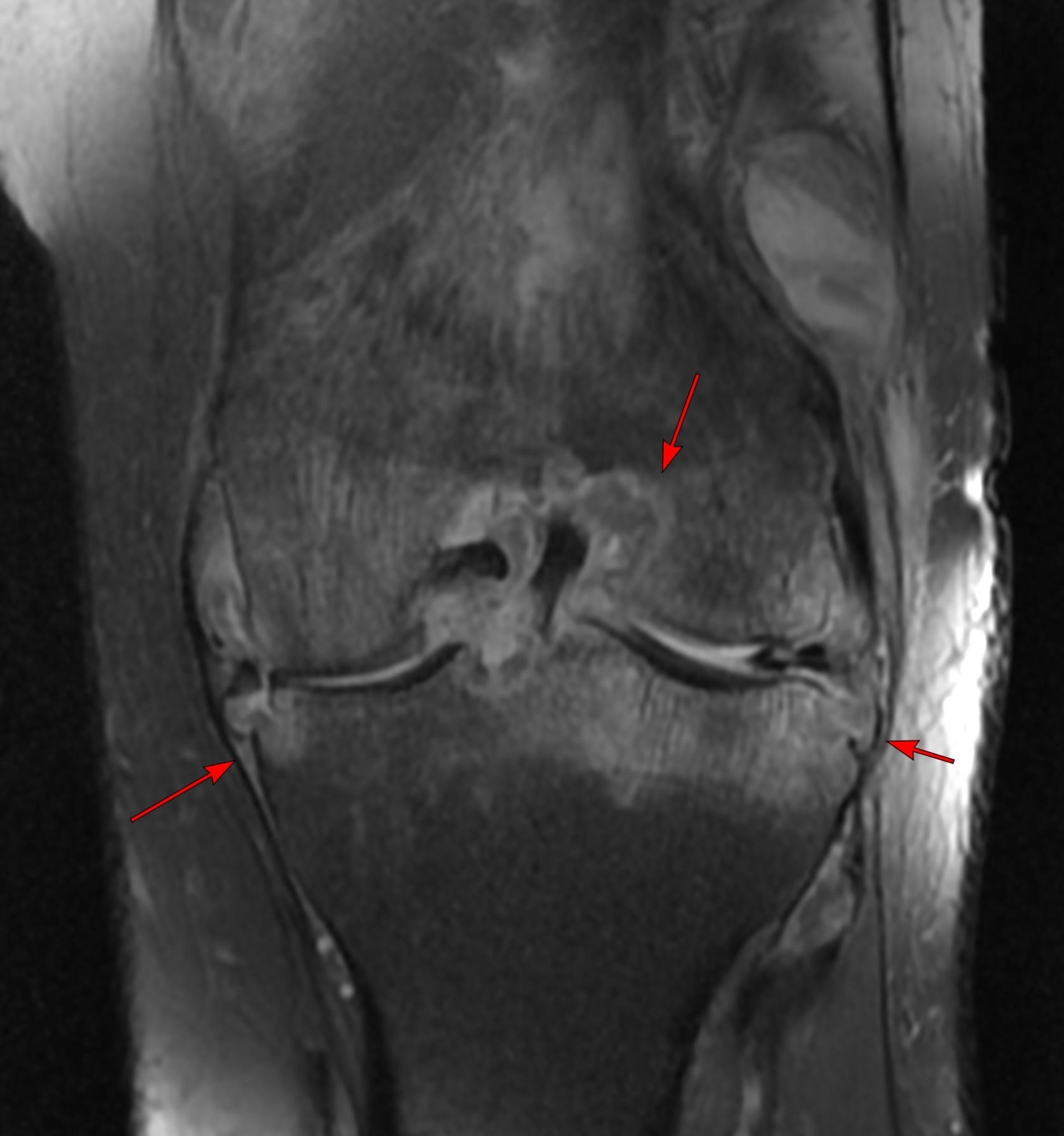
The PD fat sat coronal image (FIg. 3) shows marrow edema with free margin erosions and large central erosions (arrows).
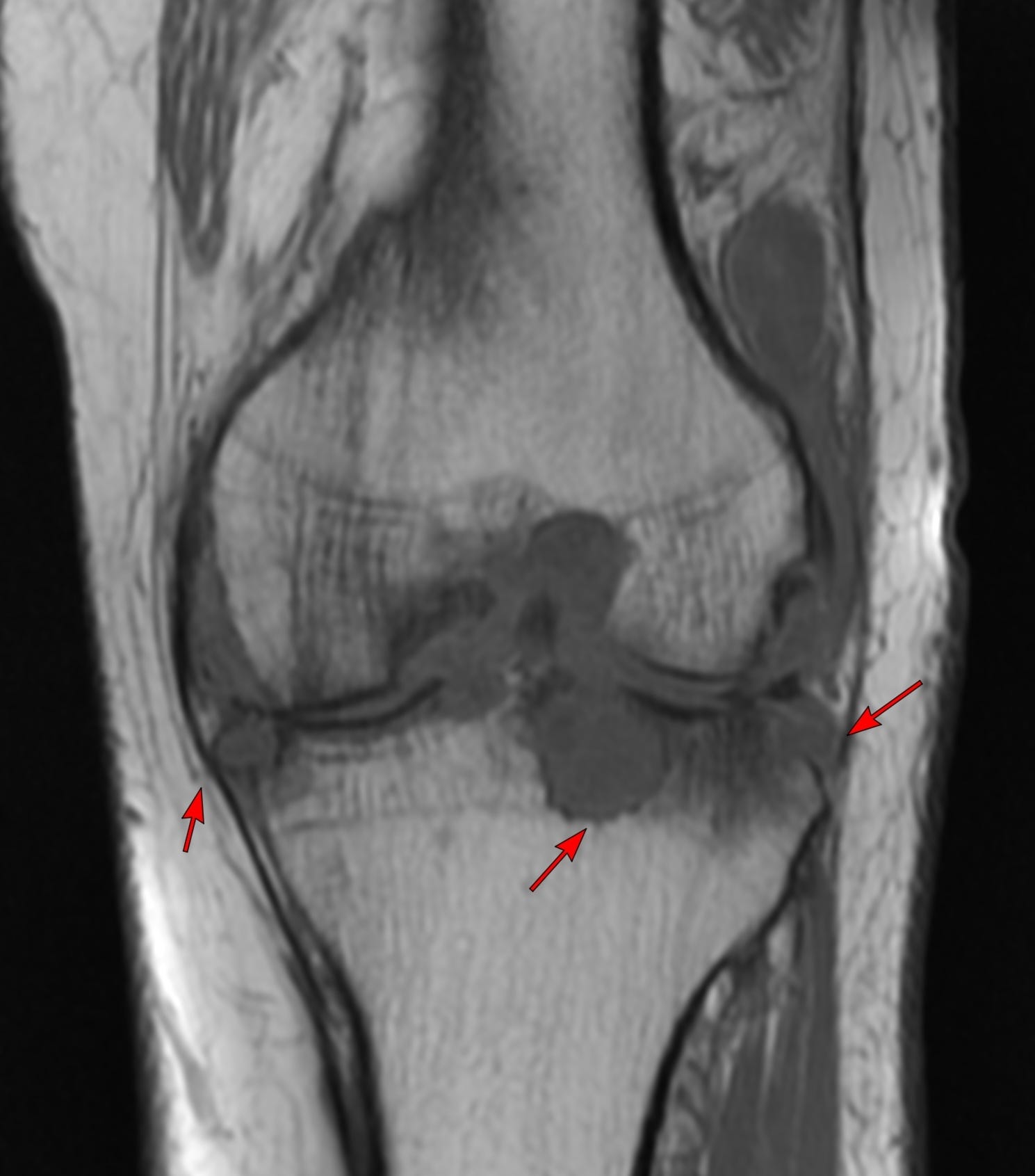
The erosions (arrows) are much better appreciated on the T1 coronal image (Fig. 4).
This is a mono-articular arthropathy. While tenosynovial giant cell tumor (see case below) may occur with T2 dark large intra-articular nodules, they rarely produce free edge erosions and marrow edema.
Case 4: Intra-Capsular Popliteal Fossa Mass

This 28-years old presented with swelling in the popliteal fossa and the inability to flex the knee fully with pain.
A mono-articular arthopathy should be assumed to be infective unless proven otherwise and this pattern is commonly seen with tuberculosis.
A biopsy confirmed the diagnosis.
This case also shows Phemister’s triad.
Other Tuberculosis Cases
Case 59: Spina Ventosa is More than Just Dactylitis

Please subscribe, if you would like to know each time a new post is published

Case 22: A Swollen Forefoot

The Updated ToCTo receive a new quiz or case once a fortnight please subscribe - it’s all free.
Index and Table of Contents

Previous Post
Case 64: The Foggy Pattern of Myositis

Please subscribe, if you would like to know each time a new post is published
Content So Far
65 Cases
16 Cases of the Day
5 Lectures