
Case 66: Solid-Variant Aneurysmal Bone Cyst (ABC)
14-years old presented with a leg swelling

Case:
14-years old presented with a leg swelling.
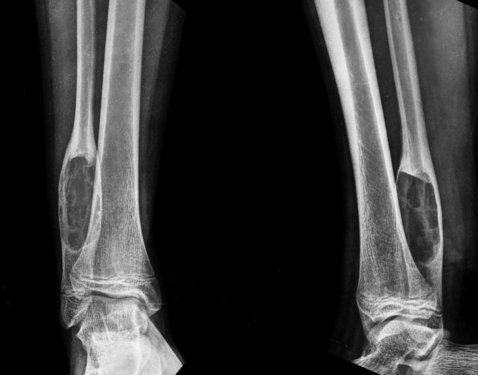
The radiograph (Fig. 1) shows an expansile, osteolytic lesion with a narrow zone of transition and a sclerotic rim, diaphyseal in location, central medullary. No marrow edema, cortical break or extra-osseous extension is seen.
This is a benign lesion. Trabeculated, expansile lesions like these in general can be
1. Non-ossifying fibroma / osteofibrous dysplasia / fibrous dysplasia
2. Giant cell tumor
3. Aneurysmal bone cyst (ABC)
4. Hemangioma
5. Desmoplastic fibroma
6. Brown tumor
The location and age rule out all except aneurysmal bone cyst. Hemangioma is very rare. The serum calcium and alk phos were normal.
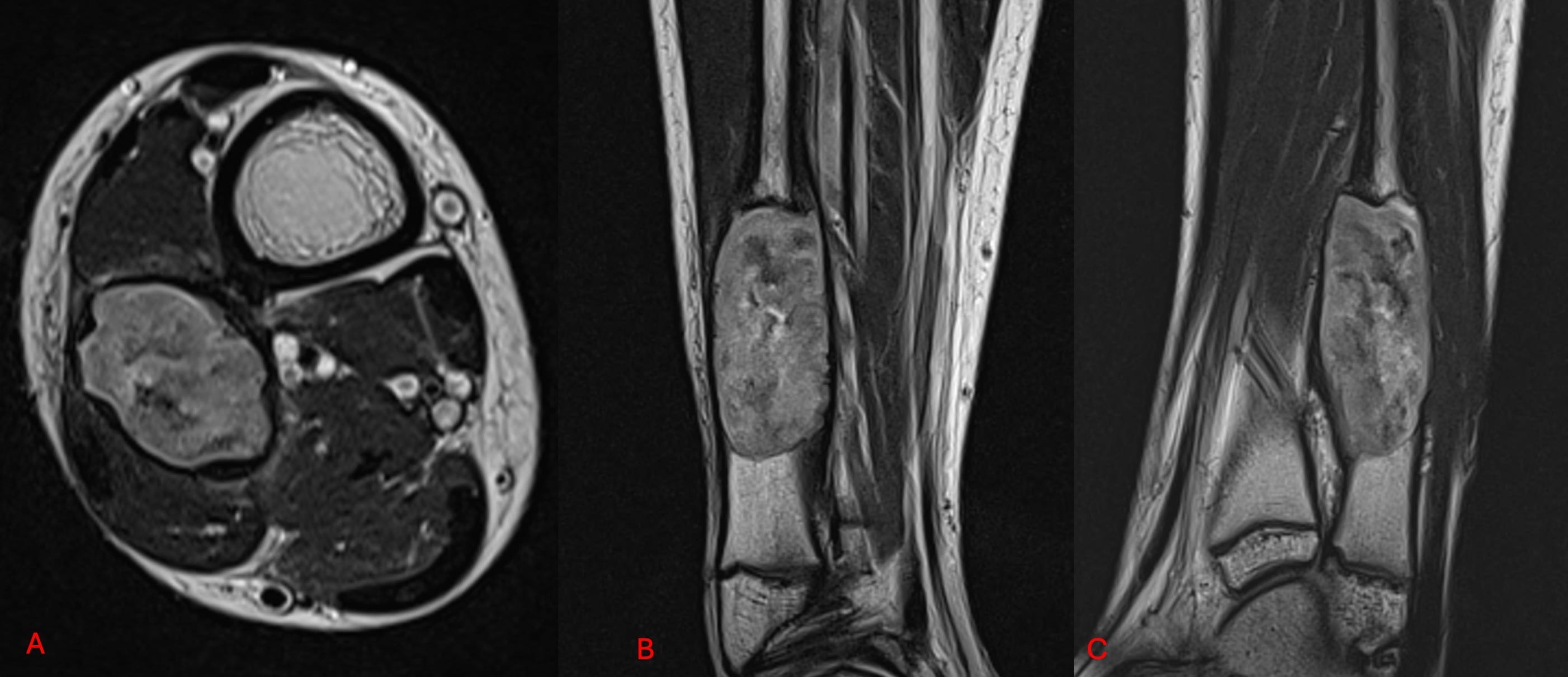
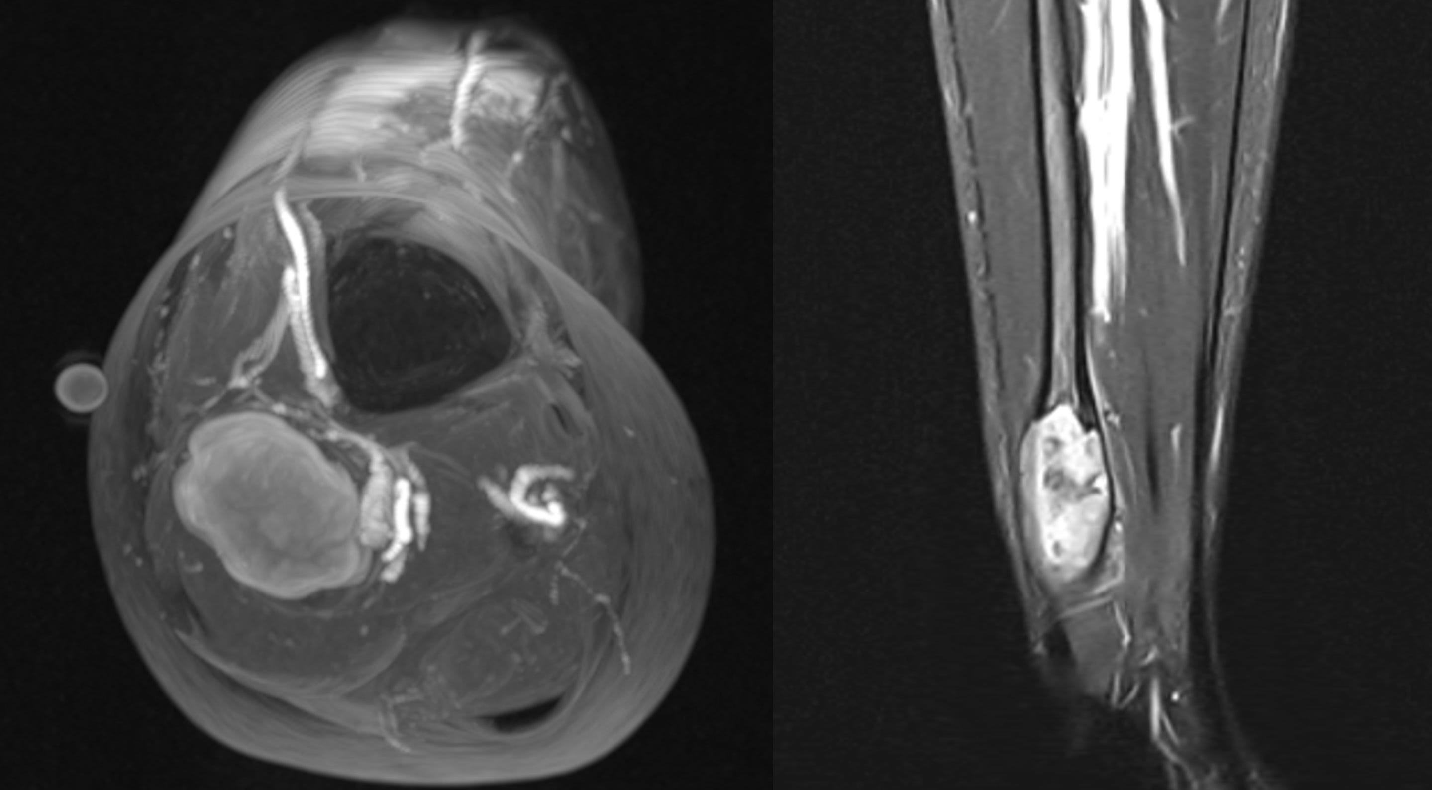
The MRI (Fig. 2) shows an expansile T2 intermediate to dark lesion (axial - A, coronal - B and sagittal - C). There are no fluid levels. The lesion is arterially enhancing (Fig. 3) lesion.
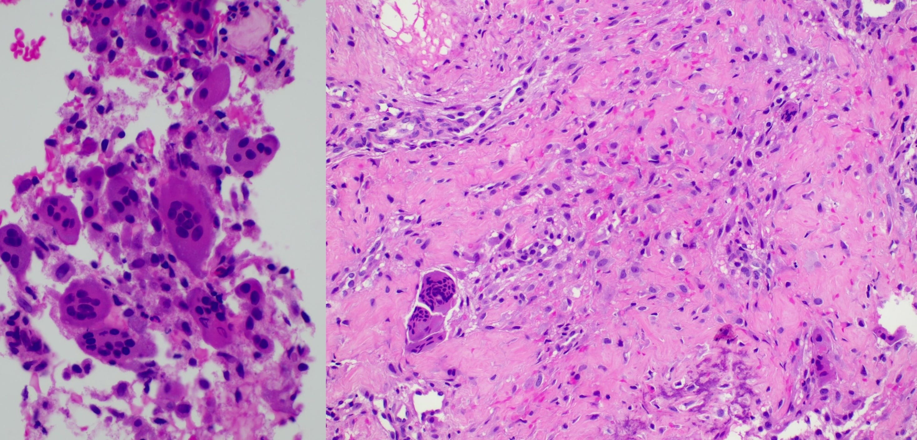
A biopsy (Fig. 4) shows a giant cell containing benign tumor.
This combination of a giant cell containing tumor with an expansile, trabeculated appearance in the diaphysis is consistent with a solid-variant ABC.
When a lesion looks like an ABC on a radiograph but does not have fluid levels and pathology shows a giant cell containing benign lesion, that is when we can make a diagnosis of solid variant ABC.
The video (YouTube Members Only) discusses the case, 2 other similar cases, the literature, then subperiosteal and surface ABCs that can also have solid variants and ends with the concept of USP6 positive neoplasms.
Other ABC Cases
Case 24: A Soapy Humerus

The Updated ToCIf you would like to receive the case in your inbox, each time a new one is up (twice a month), do subscribe
Index and Table of Contents

Previous Post
Case 65: Mono-articular Knee Arthropathy

Please subscribe, if you would like to know each time a new post is published
Content So Far
66 Cases
16 Cases of the Day
5 Lectures