

Case:
This was posted on YouTube as Post 55.
A 20-year old presented with left hip pain.
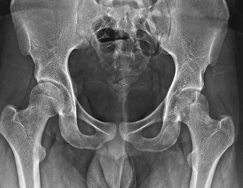
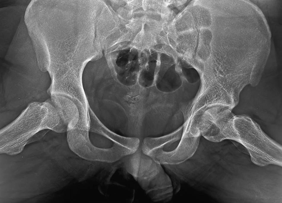
The radiograph showed a well-defined osteolytic lesion in the epiphysis of the left femoral head (Figs 1, 2). There is a sclerotic rim, which means that this is a benign/slow-growing lesion.
The MRI (Fig. 3) showed marrow edema and effusion without involvement of the acetabulum, with an epi-metaphyseal lesion, with crenated margins, with cortical thinning without a cortical break.
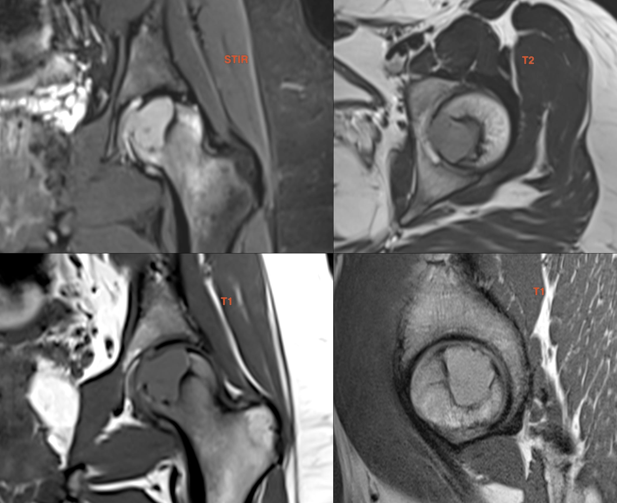
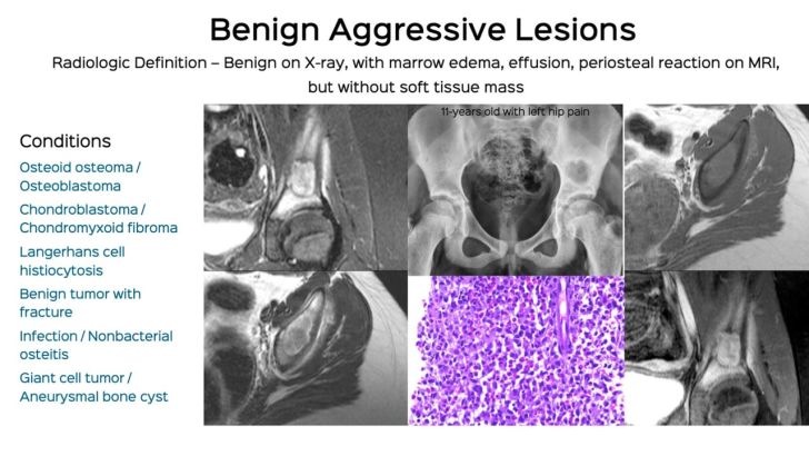
The marrow edema implies it is locally aggressive...the radiograph suggests it is benign. Hence this falls into the "benign aggressive" category.
Lecture Snippet: Benign Aggressive Lesions
If you would like an email, the moment a post is up (1-2 times a month)
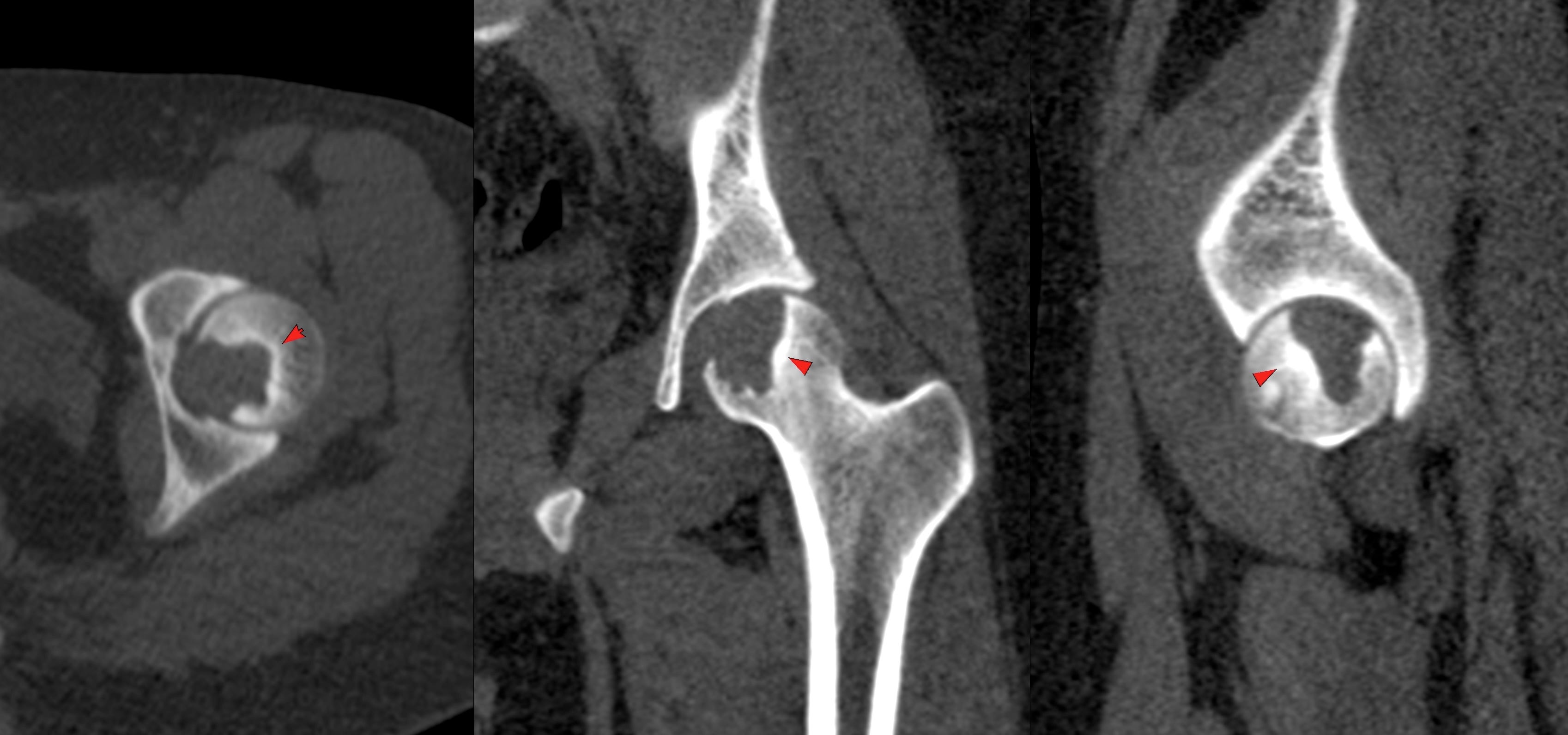
Fig. 4 shows the CT appearance. A well-defined osteolytic lesion with a sclerotic rim in the epiphysis.
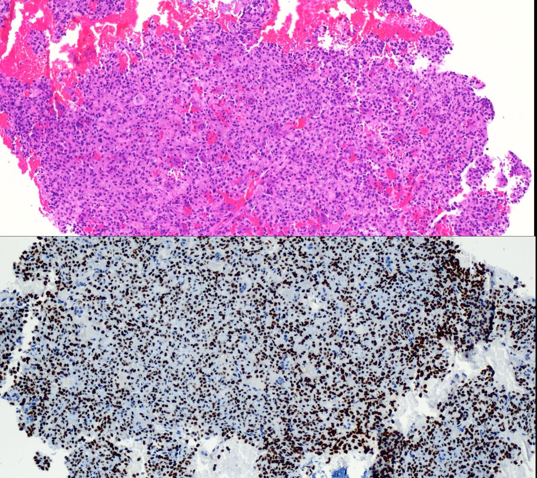
The lesion was biopsied and showed a chondroblastoma, which I also ablated using cryoablation.
This YouTube members only video discusses the case and differentials and 8 other similar cases in detail.
Other Similar Posts
Case 6: Painful Shoulder on Anti-Tuberculous Treatment

A 22-years old woman presented with a painful shoulder for 3 months. The radiographs and MRI suggested an aggressive lesion, for which she was put on anti-tuberculous treatment. When she did not improve, all the investigations were repeated. A biopsy was also then done.
Index and Table of Contents

Previous Post

Content So Far
72 Cases
16 Cases of the Day
5 Lectures