

Case:
This was also a YouTube Post No 61.
47-year-old lady presented with pain in the leg.
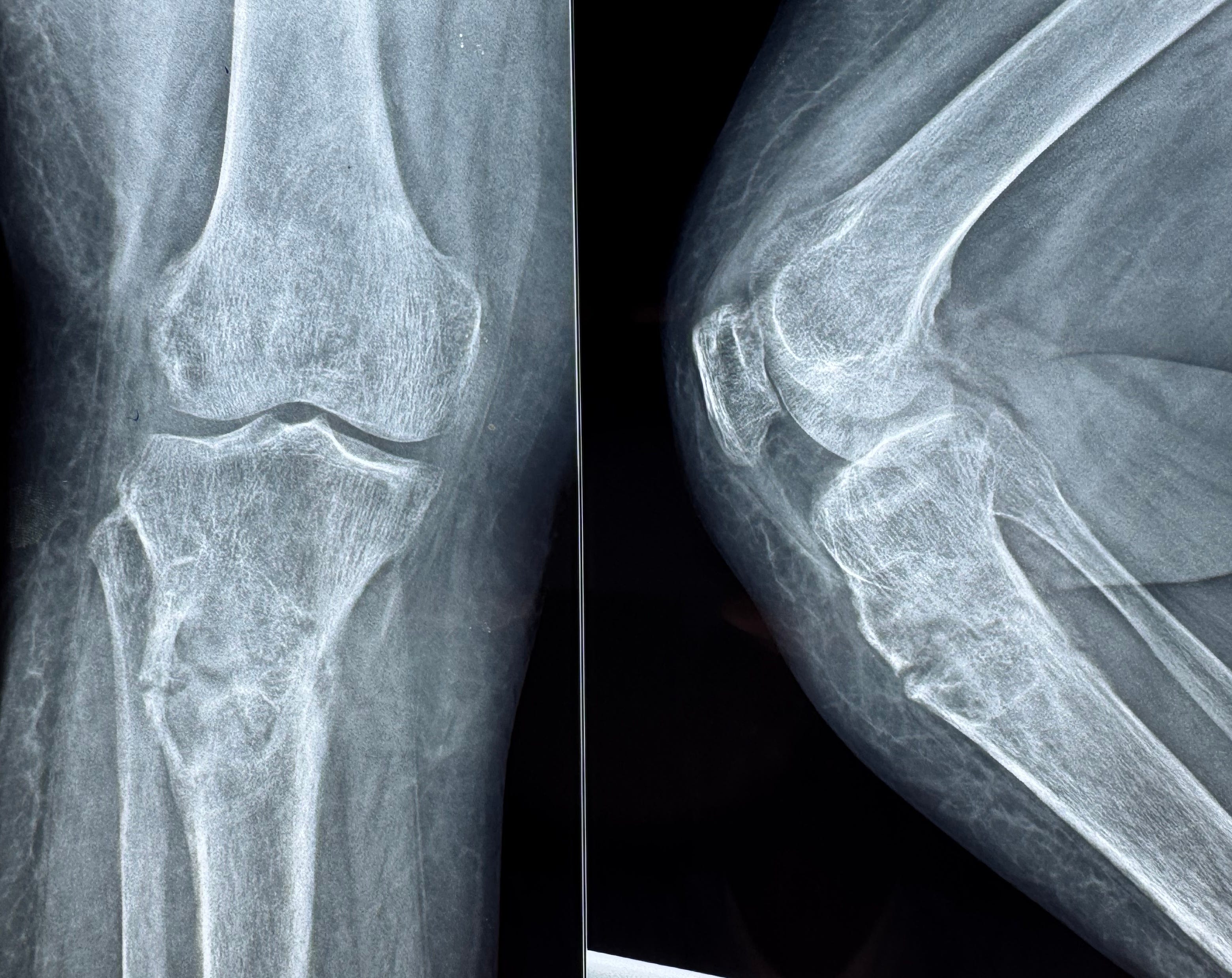
The radiograph (Fig. 1) shows an osteolytic lesion with a sclerotic rim in the proximal tibial metaphysis with a pathologic fracture.
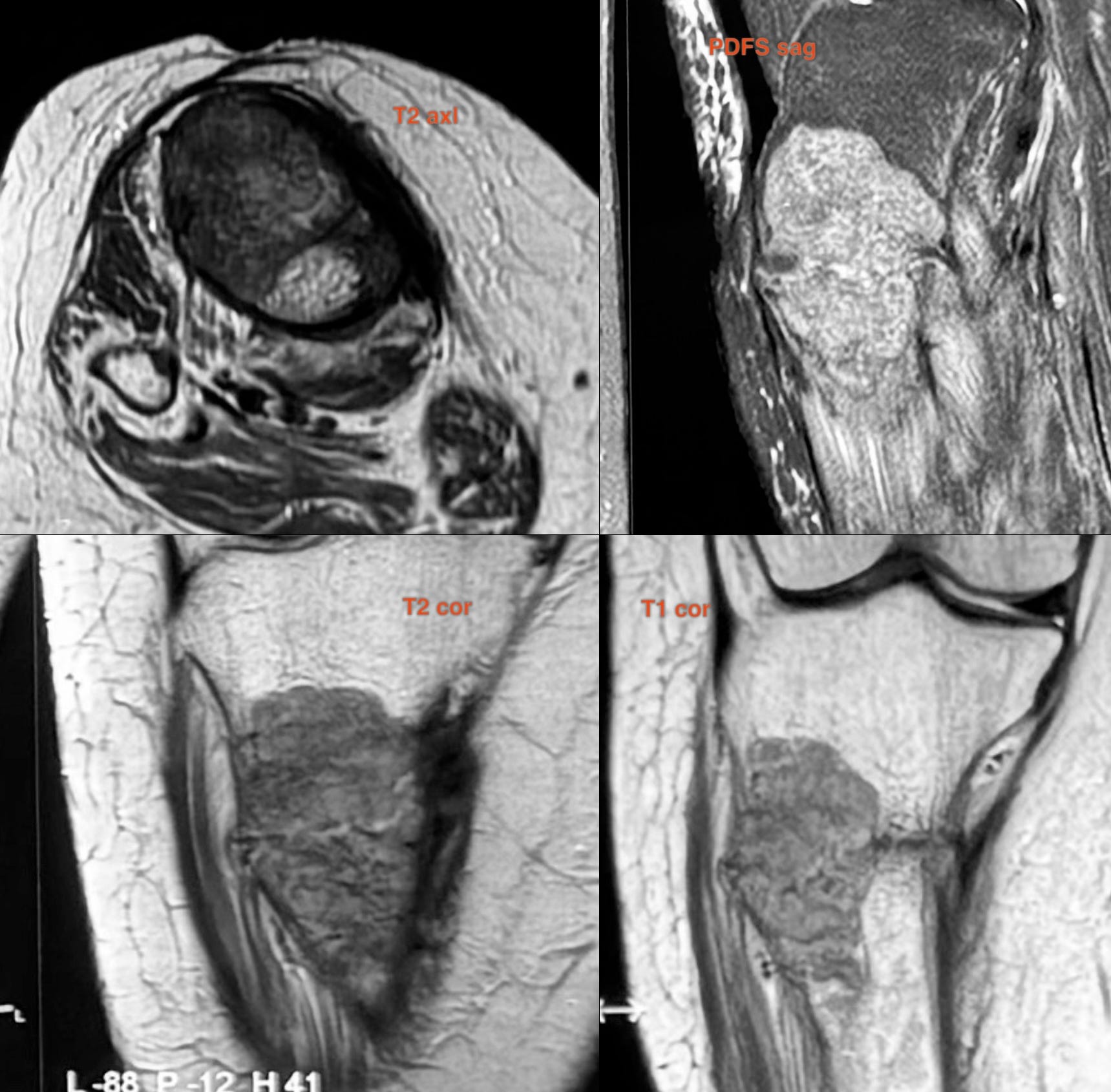
The MRI shows a T2 dark lesion with the pathologic fracture, also T1 dark with no cortical break or extra-osseous extension without marrow edema. Diffuse increased T1 marrow signal is seen with sparse trabeculae and when you go back to the radiographs (Fig. 1), the trabecular pattern is unusual.
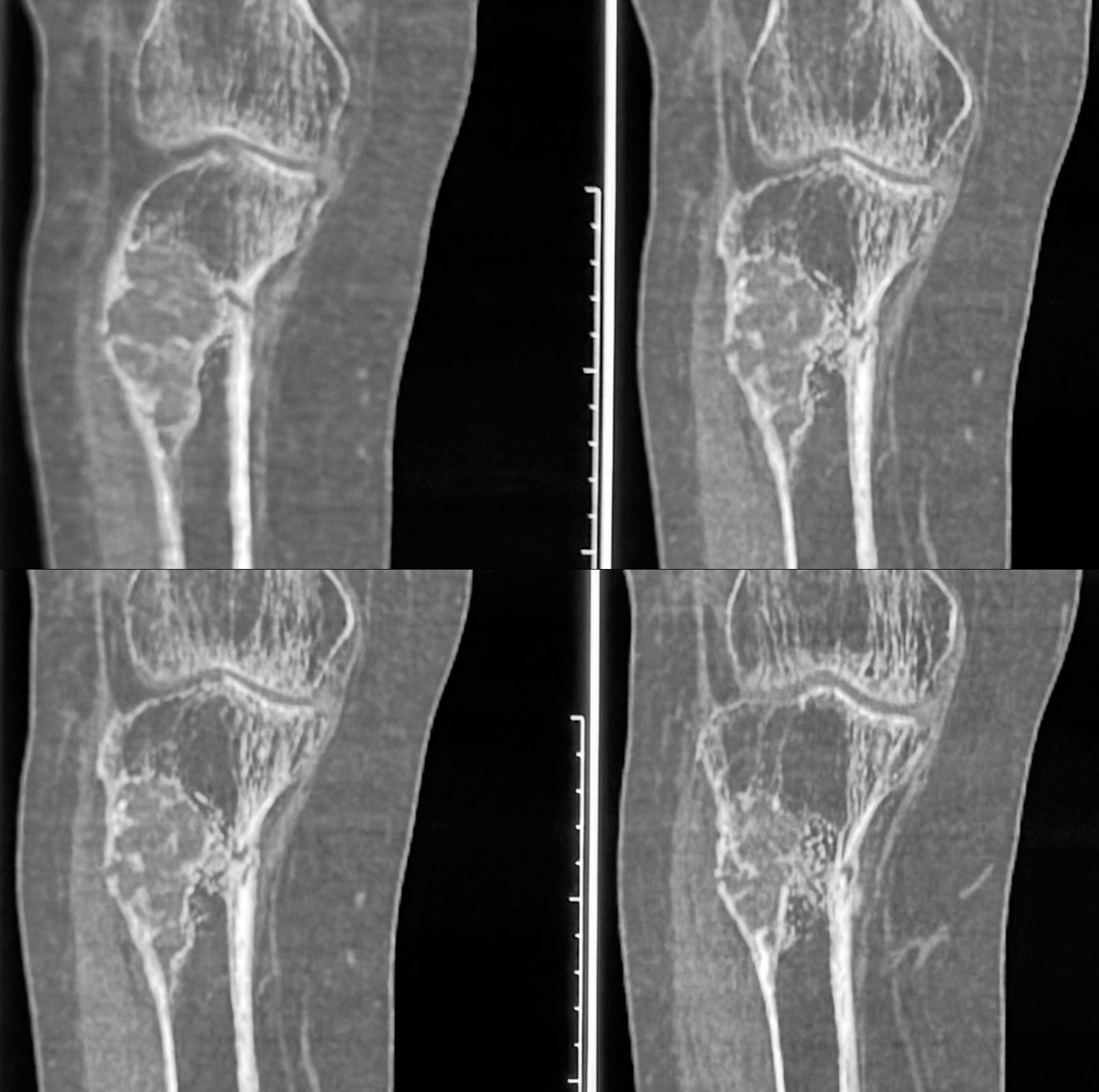
The CT scan (Fig. 3) shows a ground glass matrix with osteopenia and a markedly altered trabecular pattern.
So is this a fibro-osseous lesion? What other lesion can present with ground glass and osteoporosis?
Serum alkaline phosphatase was 886.64 U/L, normal vitamin D (36.44 ng/ml), serum calcium of 8.9 mg/dl and serum phosphorus of 2.14 mg/dl with a serum PTH of 616.4 pg/ml. So alk phos and PTH were elevated with a low serum phosphorus.
Thoughts?
The biopsy showed a giant cell containing tumor, which was histone negative.
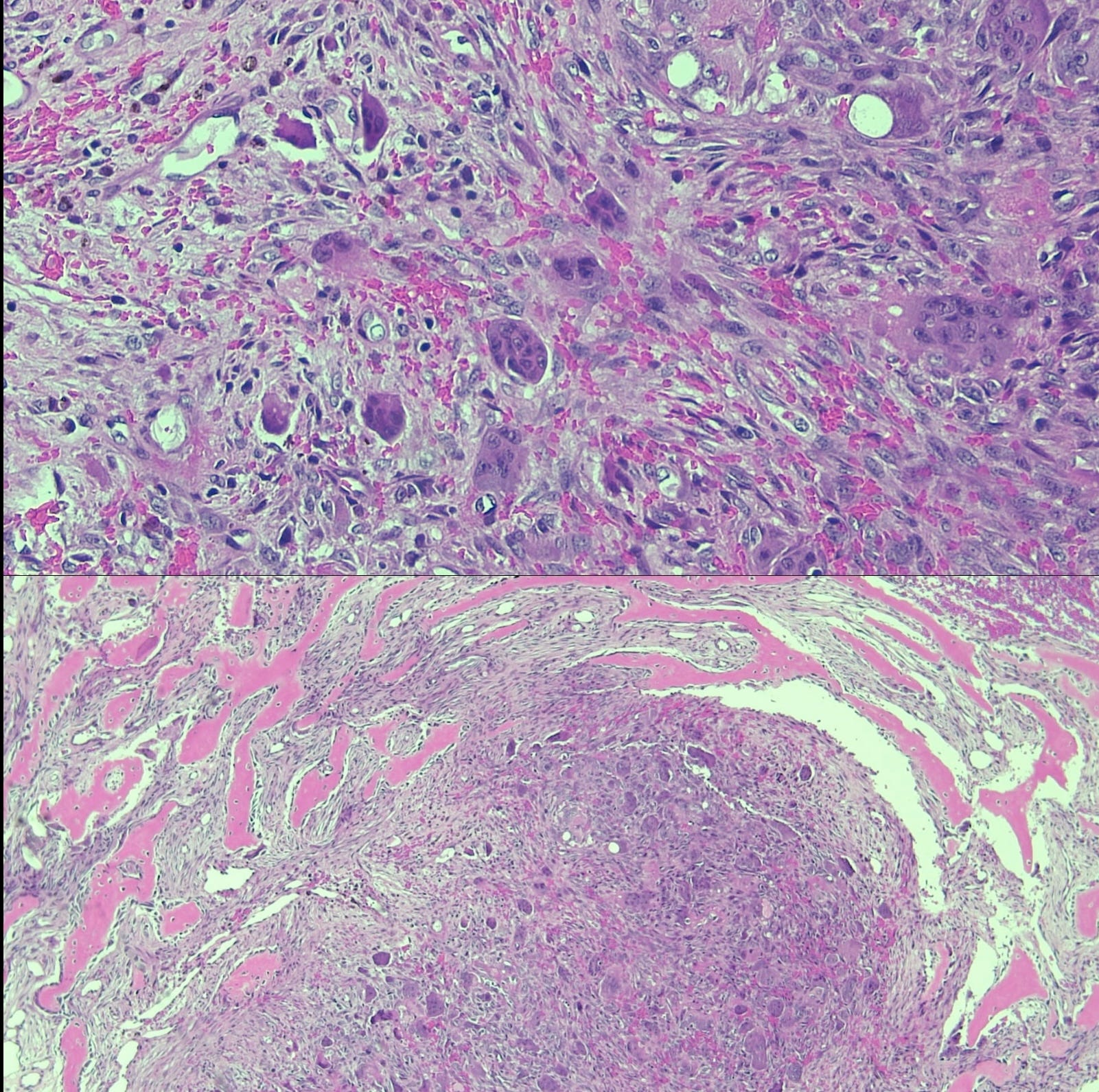
This is a classic brown tumor of hyperparathyroidism.
Other Similar Posts

Index and Table of Contents

Previous Post
Content So Far
75 Cases
16 Cases of the Day
5 Lectures