
Case 69: In Every Patient Above the Age of 35, the Most Likely Cause of an Osteolytic Bone Lesions is...
67-year-old with elbow pain

Case:
A 67-year old presented with elbow pain.
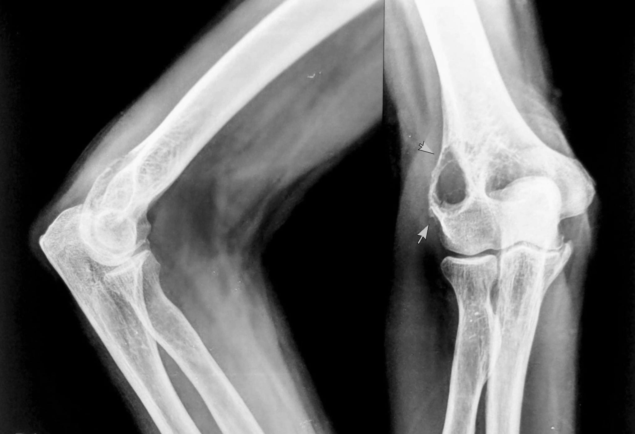
The radiograph (Fig. 1) shows an osteolytic lesion in the metaphysis involving the lateral condyle with a sclerotic rim, but with a shaggy periosteal reaction. Despite the sclerotic rim, this should be assumed to be an aggressive lesion, unless proven otherwise.
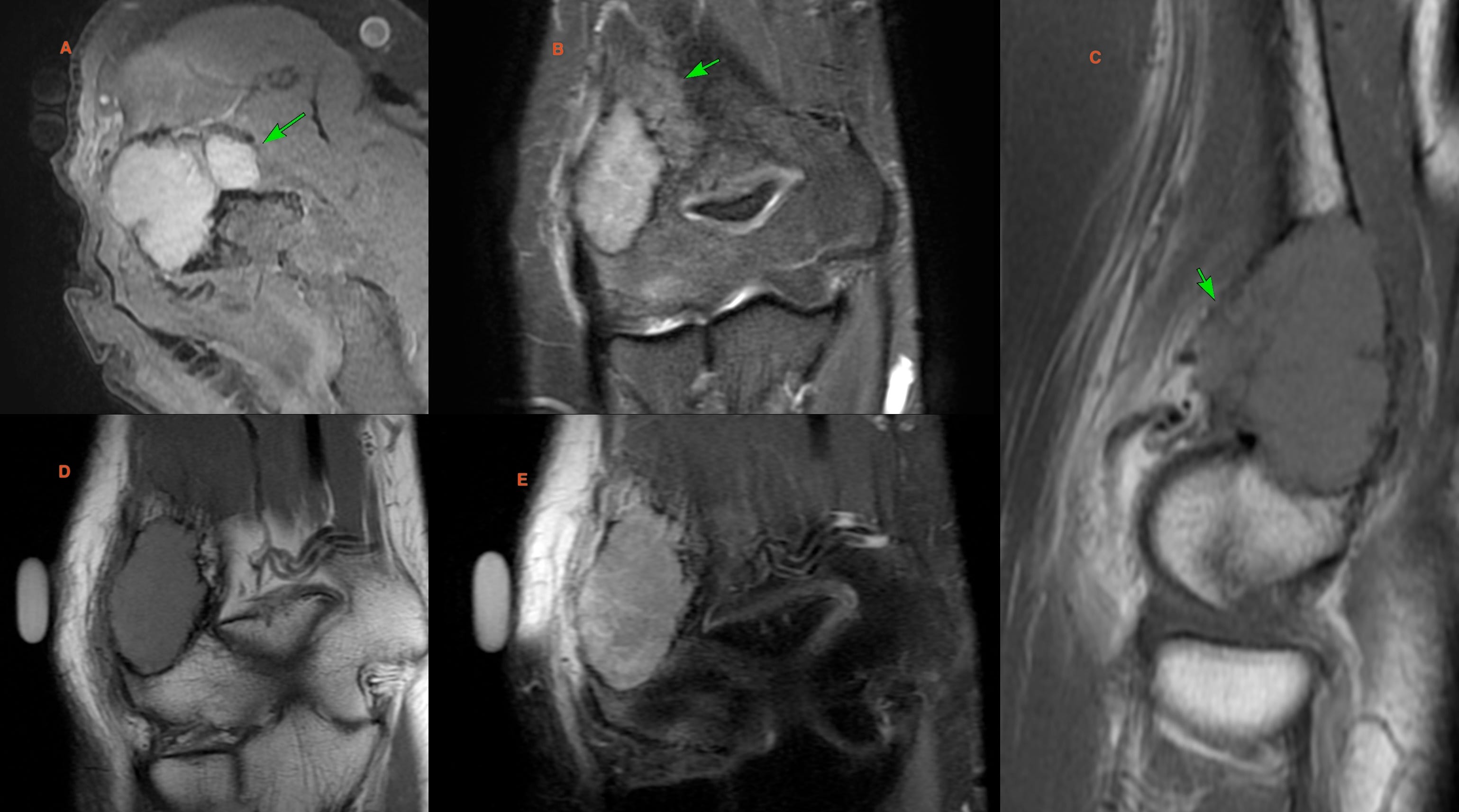
The MRI (Fig. 2) shows the osteolytic lesion with marrow edema (arrow in B) with crenated margins with extra-osseous spread anteriorly (arrows in A and C).
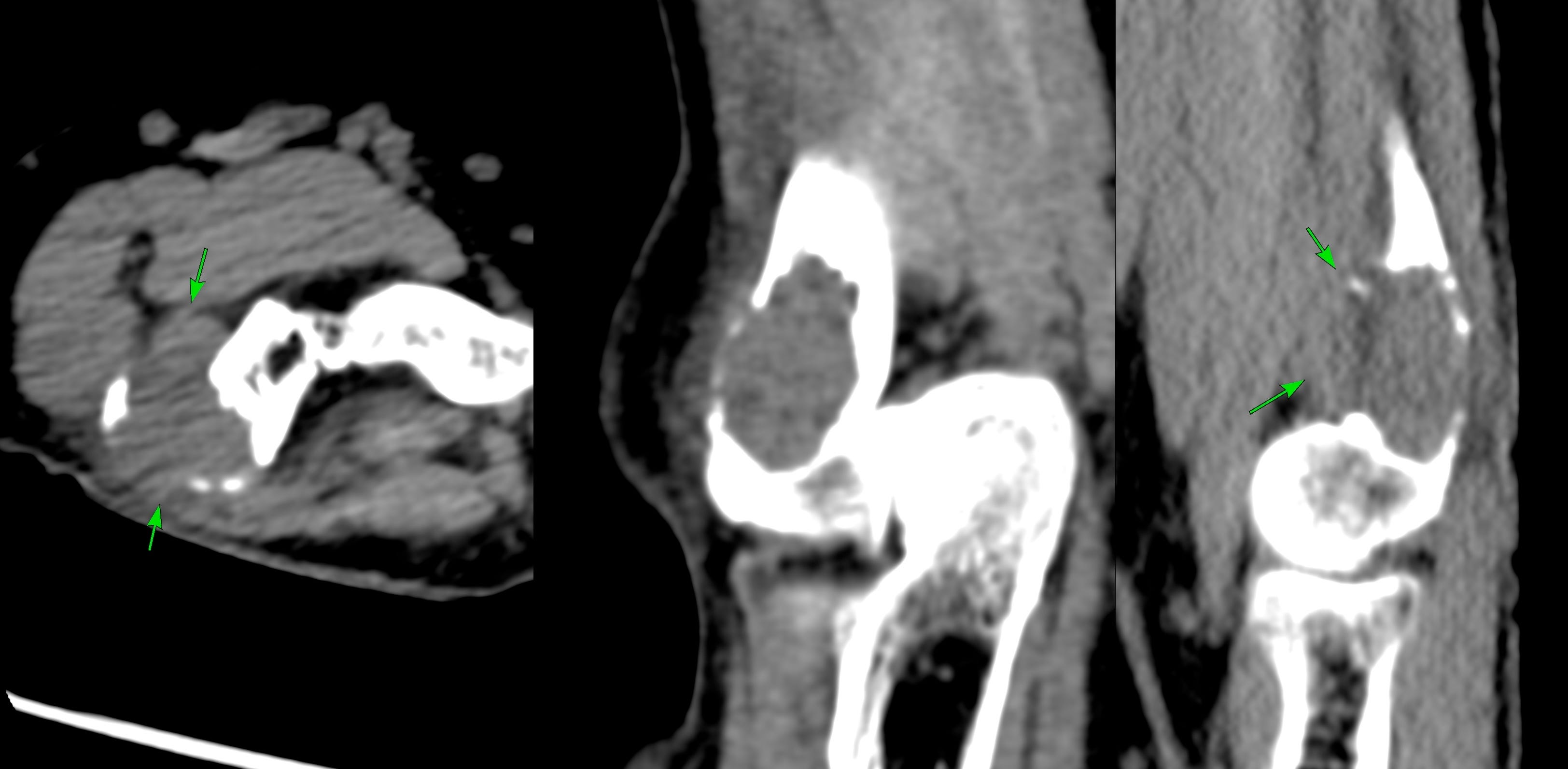
This was also confirmed on the CT scan (Fig. 3).
This is the stage where you ponder the next step. Biopsy or further imaging.
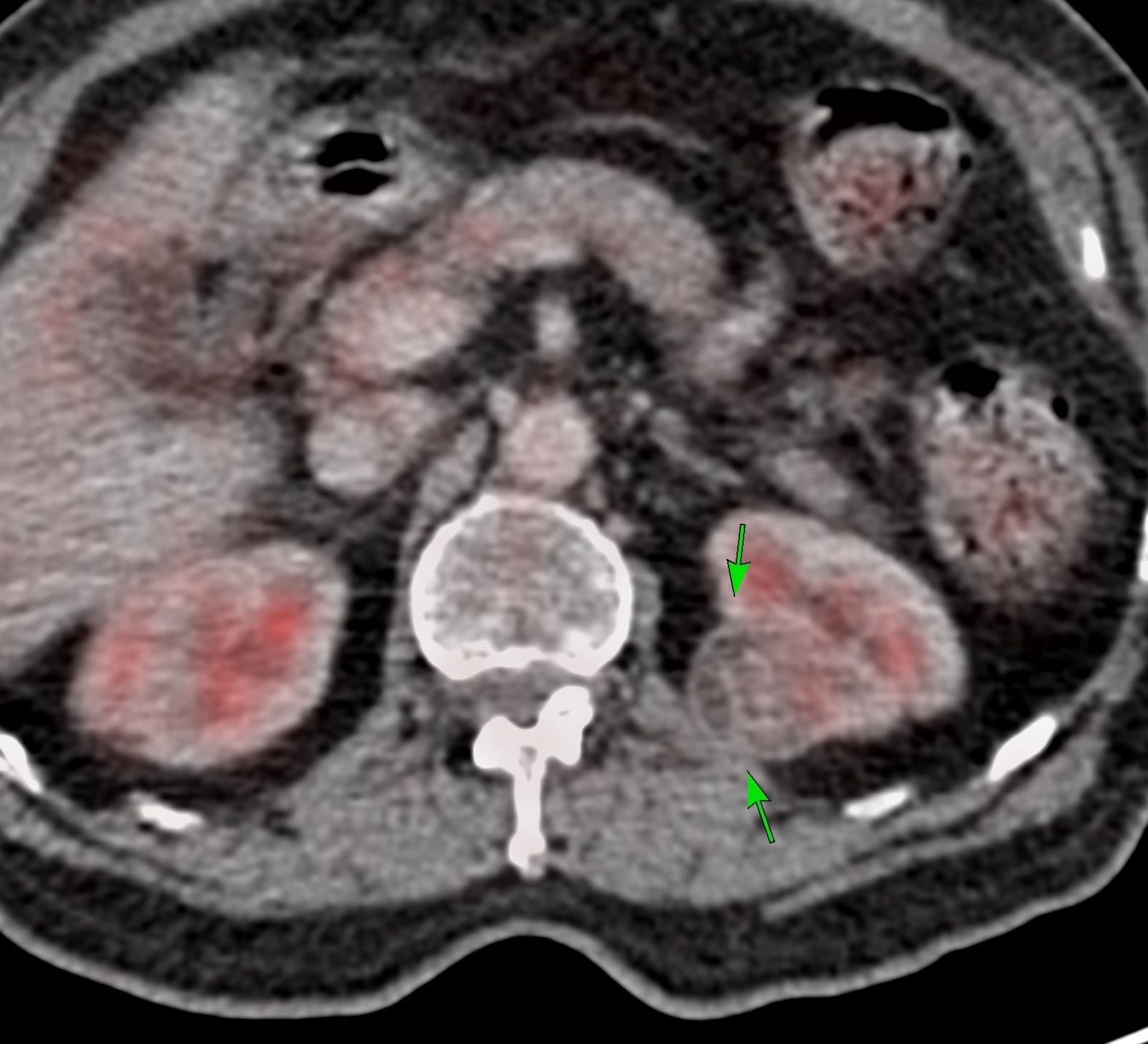
A biopsy was done and showed a clear cell malignant tumor, likely a renal primary.
A PET (Fig. 4) showed the primary lesion arising from the upper pole posteriorly.
In any person above the age of 34, unless there are specific findings pointing towards a primary bone tumor (chondroid or osteoid matrix) or infection (marrow edema, abscess, sequestrum), a focal bone lesion should be assumed to be metastasis, myeloma or lymphoma unless proven otherwise.
Last year’s lecture on Approach to Bone Tumors covered this
The first case was also about this

Other Metastases Cases
Case 37: A Sclerotic Hemipelvis

Index and Table of ContentsTo receive an email each time a new post is up (1-2 times a month)
Index and Table of Contents

Previous Post
Case 68: Identifying the Correct Matrix on Radiographs

Please subscribe, if you would like to know each time a new post is published
Content So Far
69 Cases
16 Cases of the Day
5 Lectures