

Case:
This was also a YouTube Post No 57.
22-year-old had knee pain.
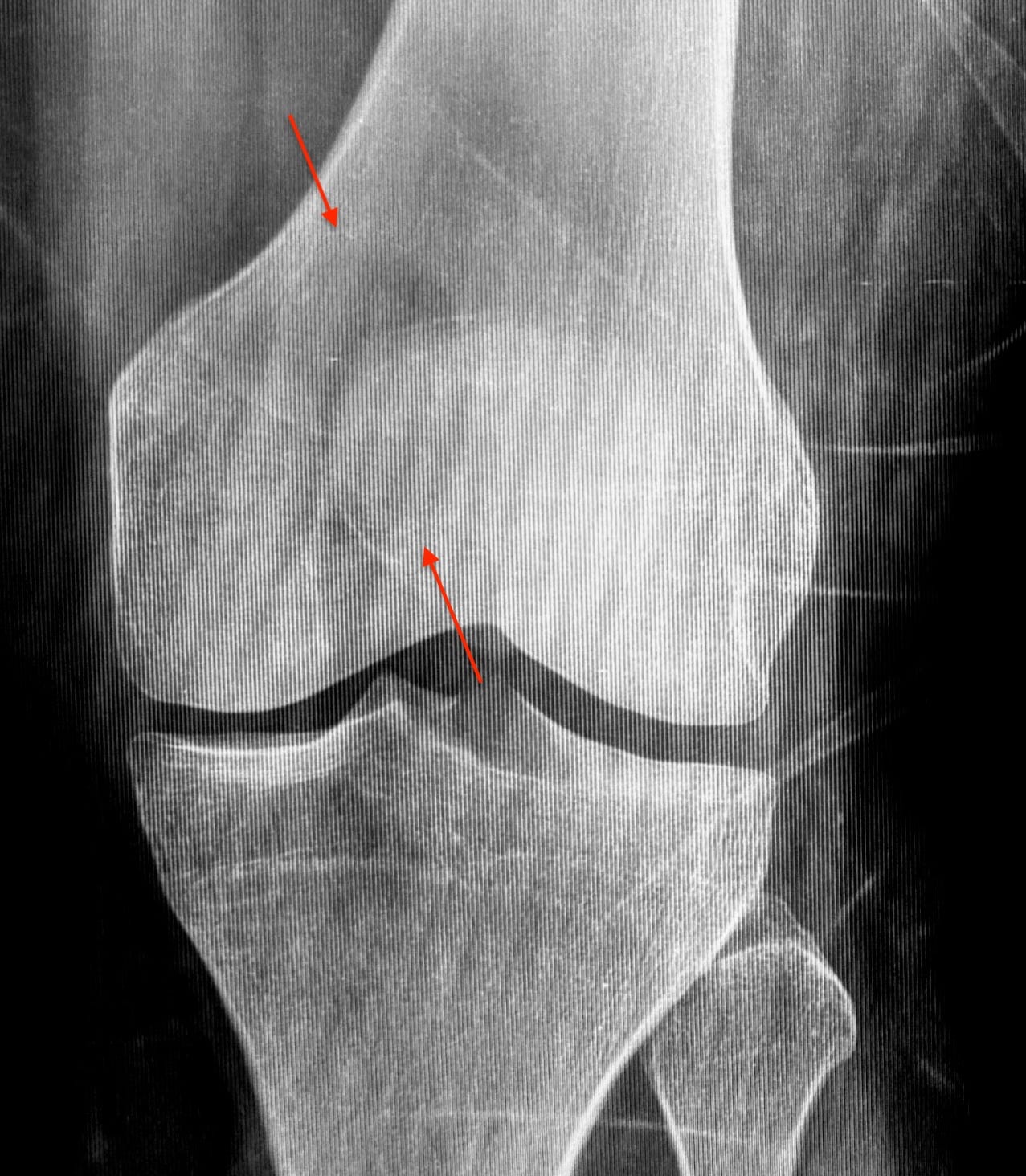
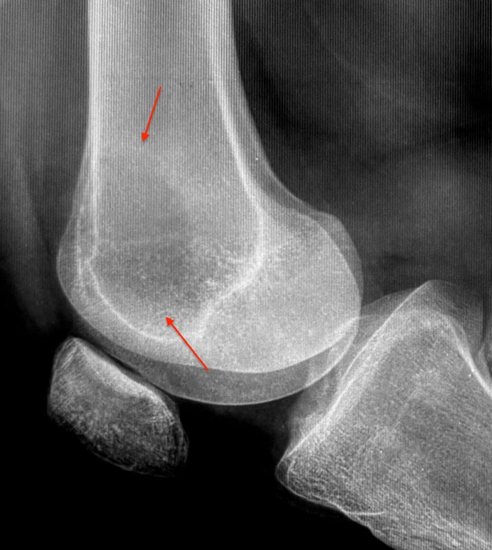
Radiographs (Figs 1 and 2) shows an osteolytic lesion with a narrow zone of transition but without a sclerotic rim in the distal femur involving the medial femoral condyle, not reaching up to the distal articular surface.
MRI (Fig. 3) shows a T2 intermediate to dark lesion with cystic areas within, epimetaphyseal, juxtamedullary without a cortical break or extra-osseous extension, with marrow edema. It shows crenated margins. The lesion does not extend to any articular surface, including trochlea.
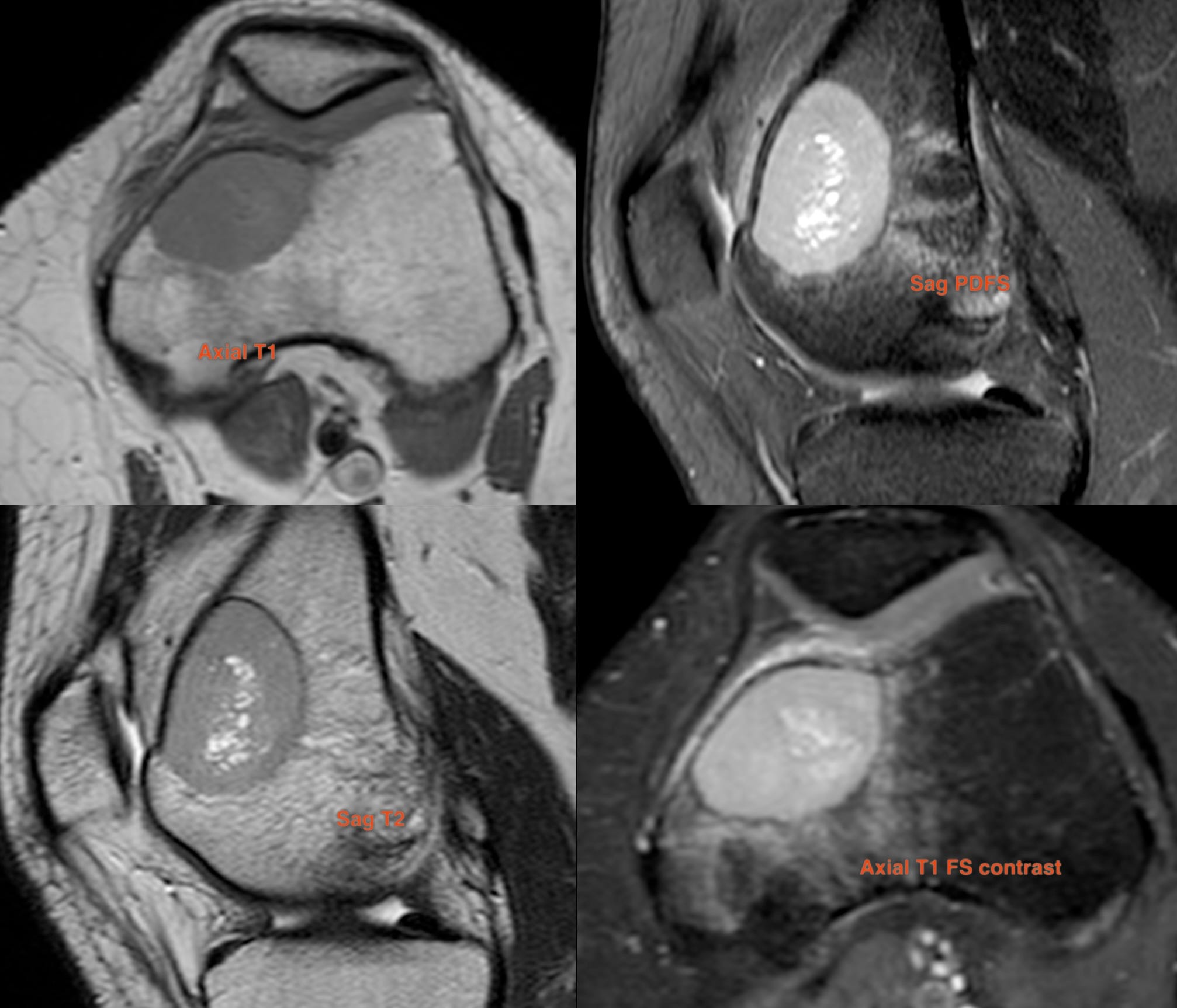
It is a likely benign lesion (but not absolutely definite, because there is no sclerotic rim) with marrow edema, so could be labeled as “benign aggressive lesion”.
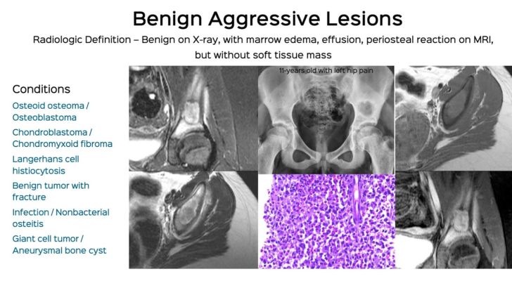
Lecture Snippet: Benign Aggressive Lesions
If you would like an email, the moment a post is up (1-2 times a month)
These are typically
osteoid osteoma,
osteoblastoma,
chondroblastoma,
chondromyxoid fibroma,
Langerhans cell histiocytosis,
benign tumor with fracture,
infection/CNO and
GCT/ABC.
Giant cell tumor (GCT) typically extends up to the articular surface, which this lesion doesn’t. Chondromyxoid fibroma (CMF) can be eccentric. None of the others fit. He is skeletally mature.
A biopsy is mandatory.
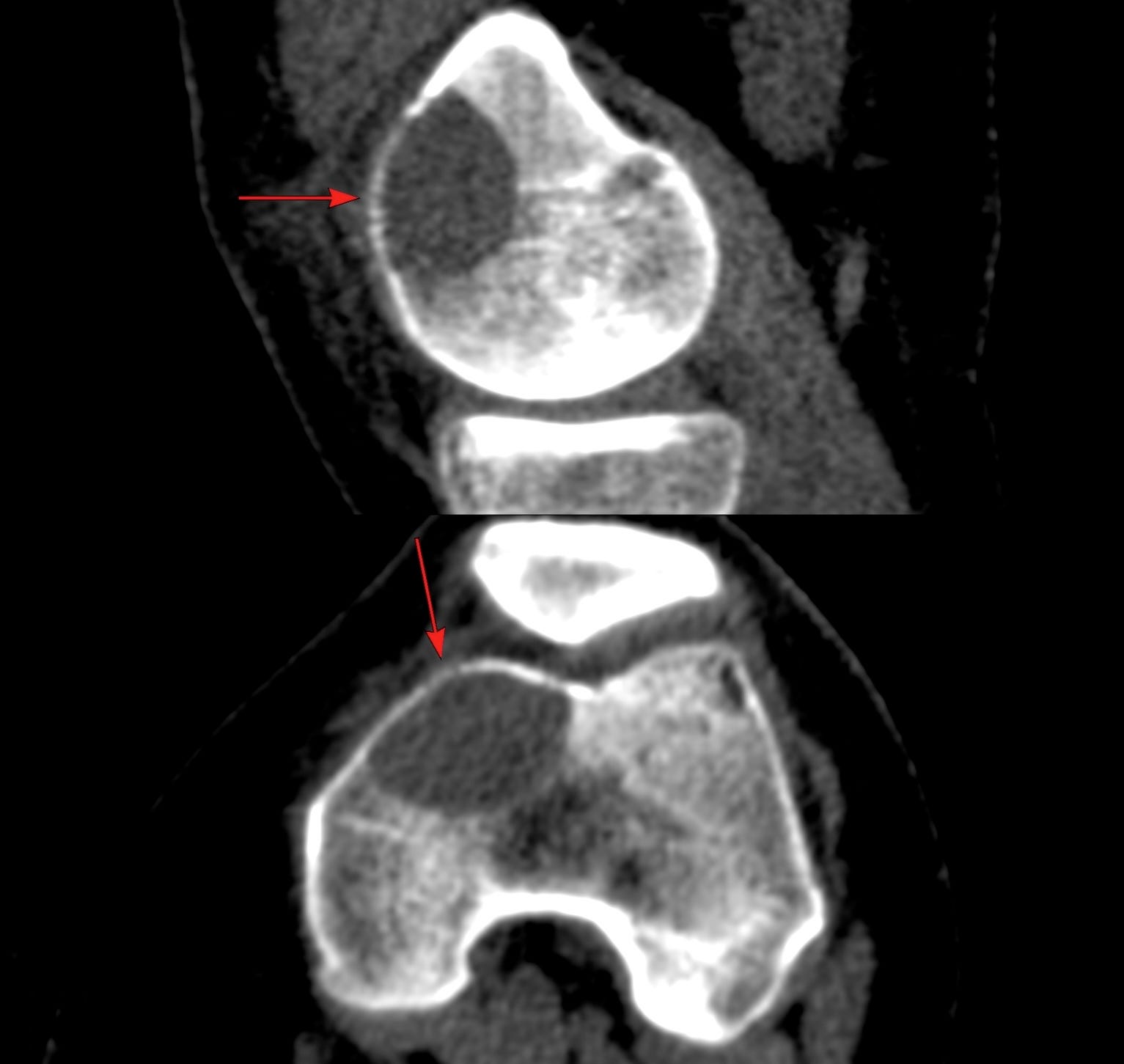
The CT scan (Fig. 4) before the biopsy shows a well-defined lesion, but without a sclerotic rim, with crenated margins (arrows). No extra-osseous spread or cortical break is seen. Crenated margins are more common with GCT, but may also be seen with malignant tumors.
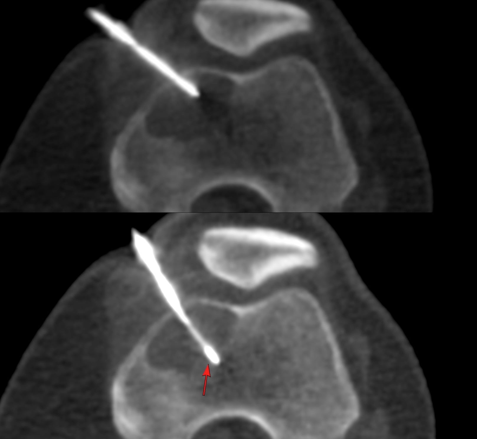
A biopsy was performed after infiltrating the periosteum with an option to give intravenous sedation if it pained. A 16G BARD needle was introduced with a light push through the cortex using an anteromedial approach (Fig. 5). Seven cores were obtained for histopath and aspirate was sent for cytology and microbiology. It took 7 mins.
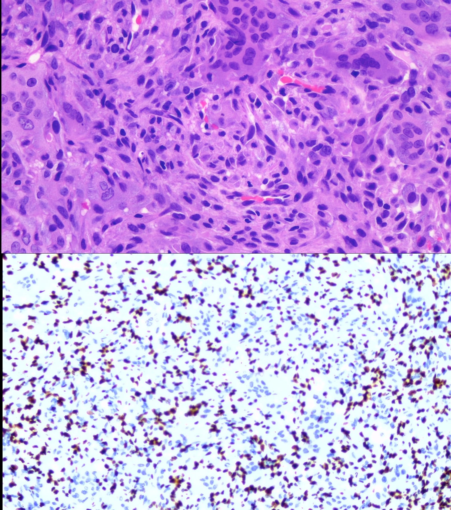
The histology (Fig. 6) shows a classic giant-cell containing tumor with was histone (H3.3 G34W) positive.
The patient was operated with curettage and cementing.
Not all GCTs extend up to the articular surface (around 1-16%) and when they don’t, it is difficult to be definite about the diagnosis without a biopsy. In any case, this is a lesion that needs a biopsy, irrespective of the pre-test possibility, which in any case is a neoplasm and not infection.
Other Similar Posts
Case 12: Typical Distal Femur Lesion

If you would like to know immediately, each time a new case is up via email, please subscribe.

Case of the Day 100 - 2025 09 10 - T2 Dark Thoracic Spinal Vertebral Lesion - Diagnosis?
Please subscribe, if you would like to know each time a new post is published
Index and Table of Contents

Previous Post

Content So Far
74 Cases
16 Cases of the Day
5 Lectures