

Case:
This was also a YouTube Post No 63.
22-year-old with swelling around the proximal wrist/distal forearm.
Radiographs shows a large osteolytic lesion with a sclerotic rim in the epimetaphysis of the distal radius (red arrows) showing cartilage. An exophytic soft tissue nodule with cartilage (rings and arcs at the periphery) is seen (yellow arrow) extending radial and volar to the bone lesion.
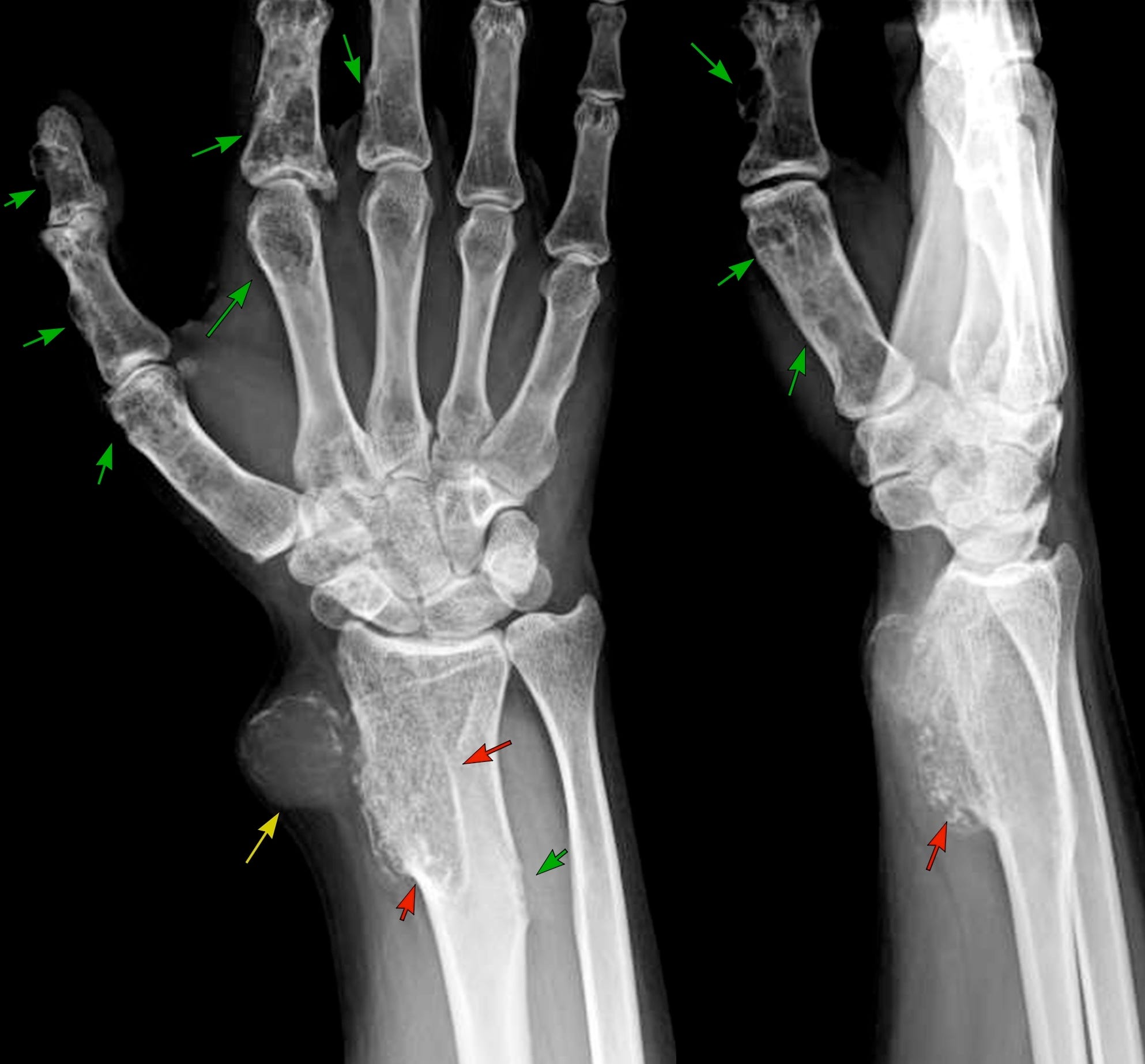
Also seen are multiple other lesions in the phalanges, all representing expansile lesions, likely cartilage tumors (green arrows), as well as in the distal radius medially.
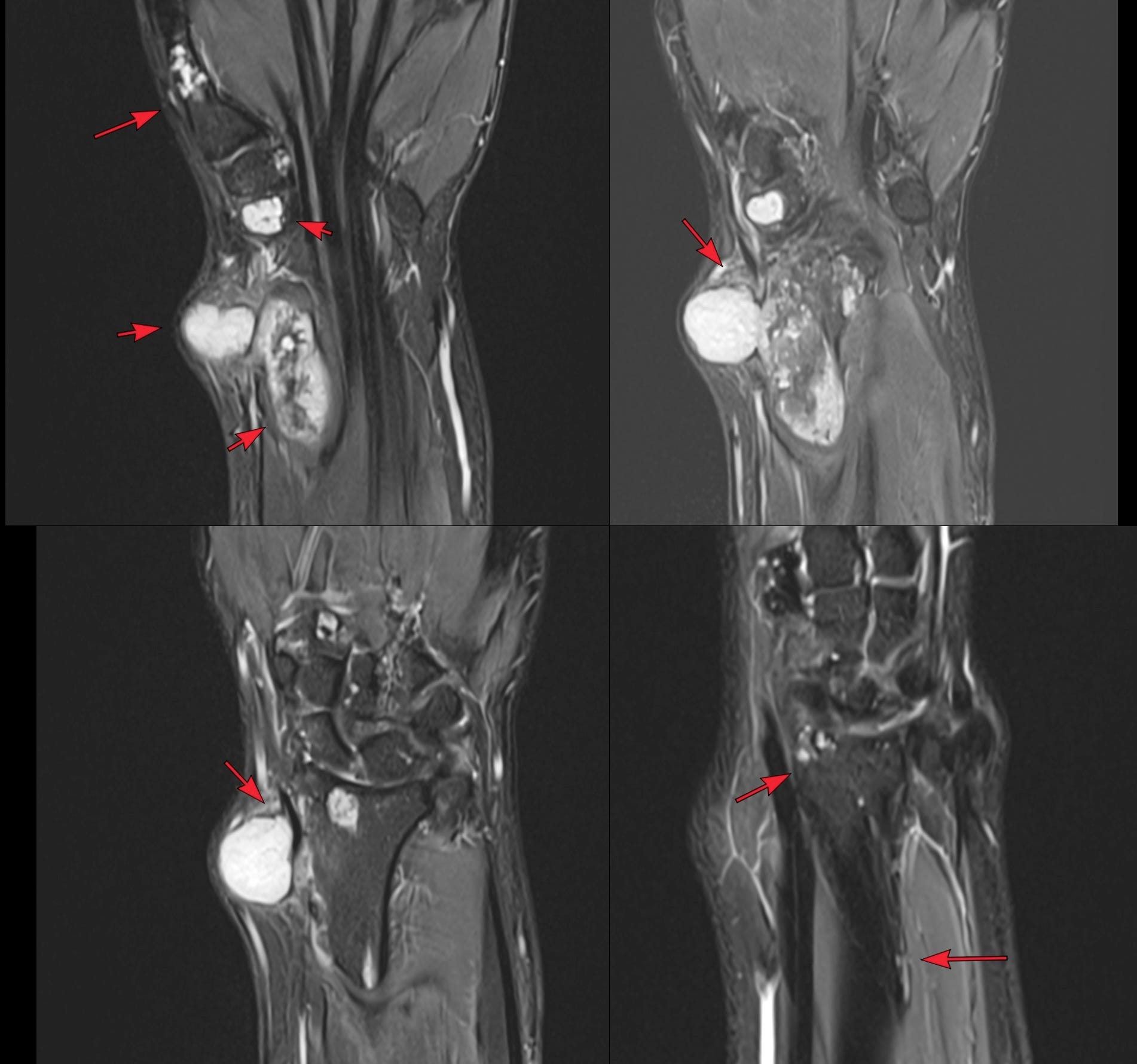
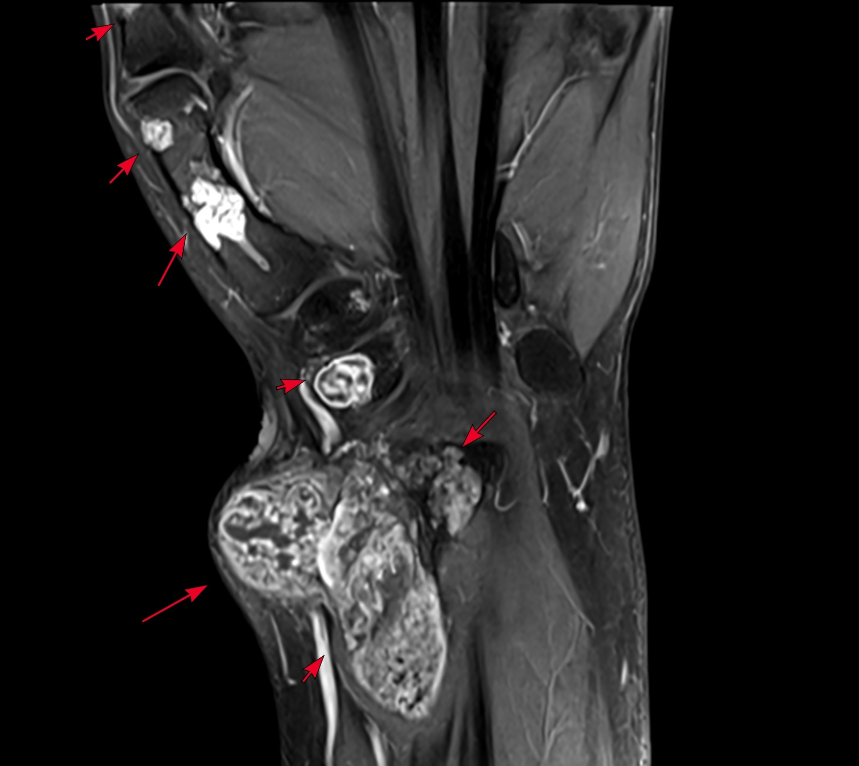
The coronal STIR MRI (Fig. 2) from volar to dorsal shows the surface and intra-medullary lesions (red arrows), along with the exophytic component.
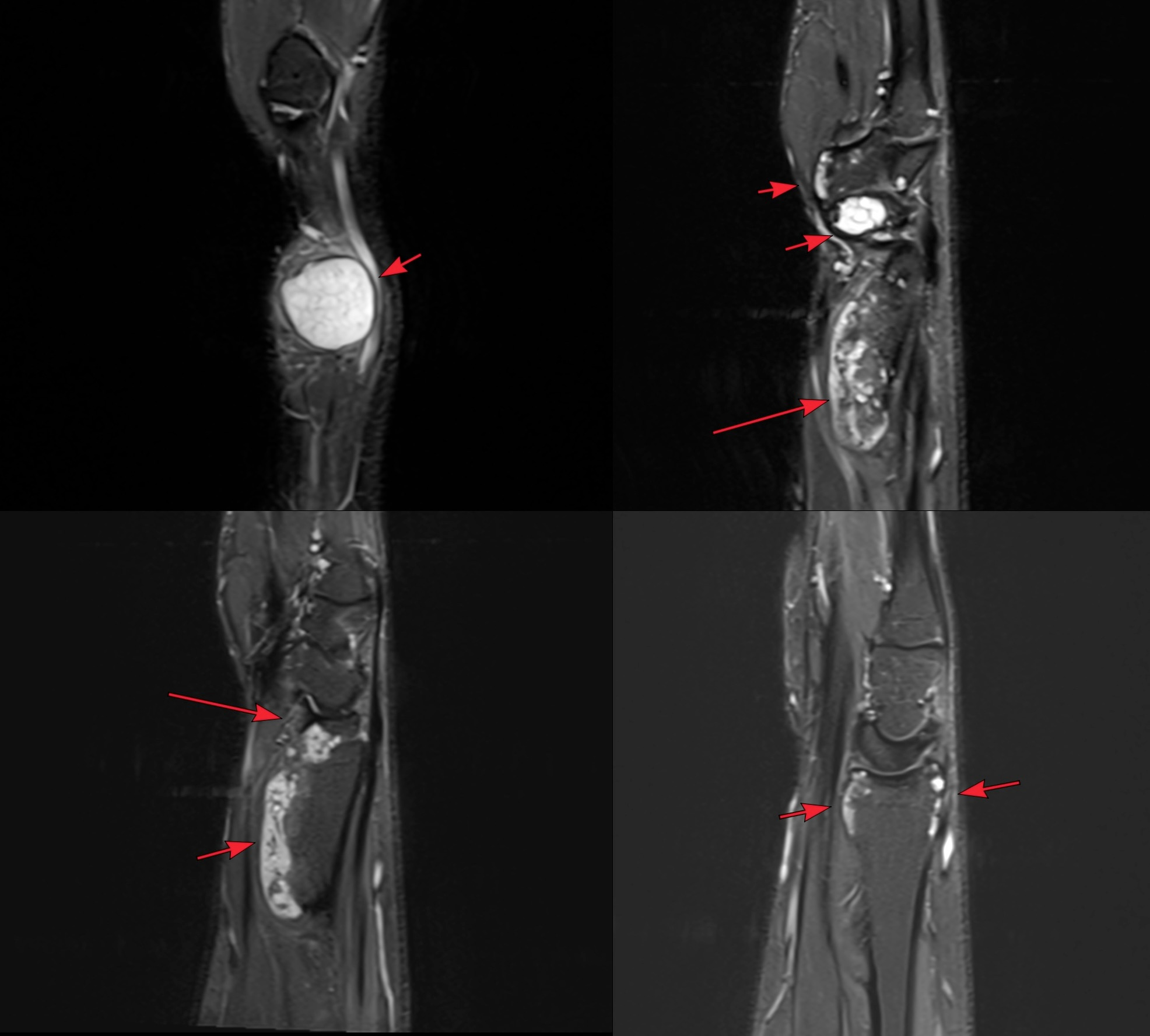
The sagittal STIR MRI (Fig. 3) from radial to midline shows the exophytic component and the rest of the intra-medullary and surface cartilage foci (red arrows).
The axial post-contrast MRI (Fig. 4) shows the large exophytic component and the rest of the surface and intra-medullary lesions with the typical pattern of cartilage enhancement.
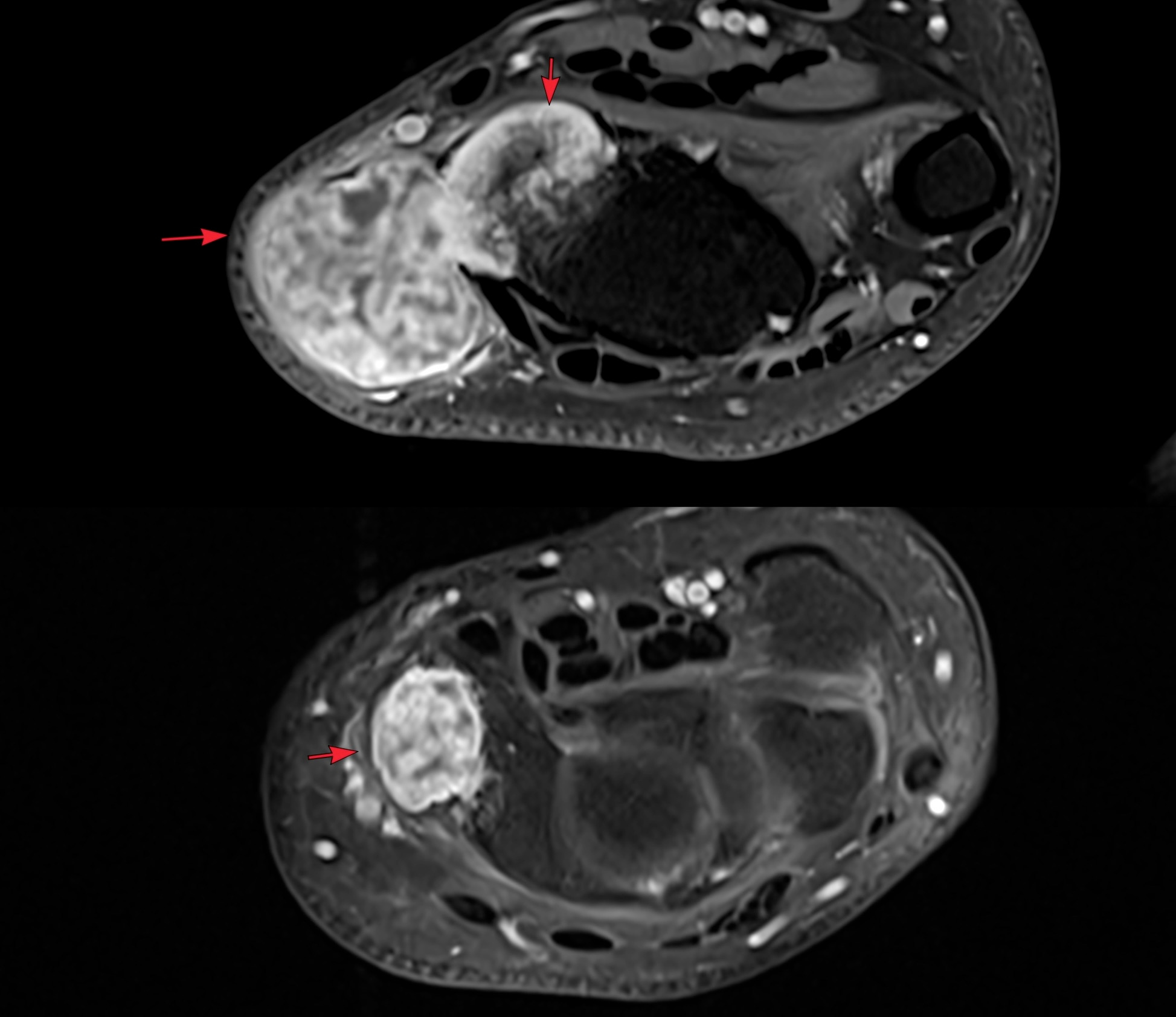
Fig. 5 is a coronal post-contrast image that shows multiple radial sided cartilage lesions.
The exophytic lesion was biopsied and the histopath (FIg. 6) shows myxoid change with changes of chondrosarcomatous transformation.
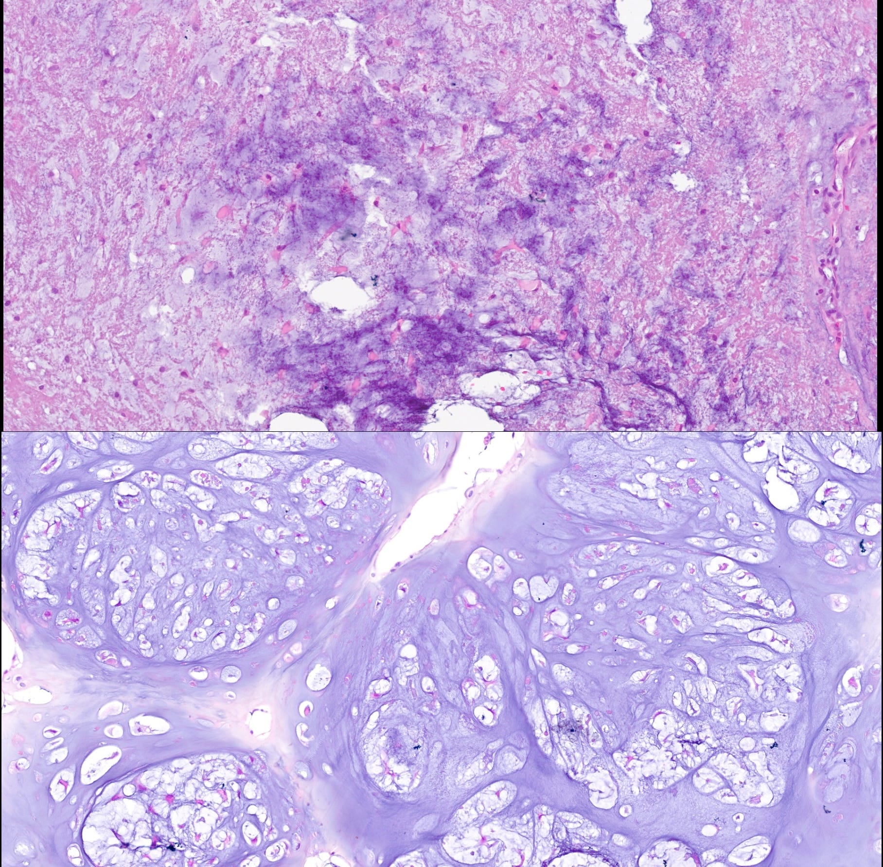
This pattern of cartilage involvement is typical of Ollier’s disease. The rate of chondrosarcomatous change in patients with Ollier’s disease is higher than in the normal population.
Other Similar Posts

Case 67: Osteochondroma or Chondrosarcoma?

Please subscribe, if you would like to know each time a new post is published
Index and Table of Contents

Previous Post
Content So Far
77 Cases
16 Cases of the Day
5 Lectures